STURGEON GERM PLASM SEPARATED FROM OTHER GERM LINEAGES AS A UNIQUE MODEL FOR STUDYING PRIMORDIAL GERM CELLS IN FISH
Introduction
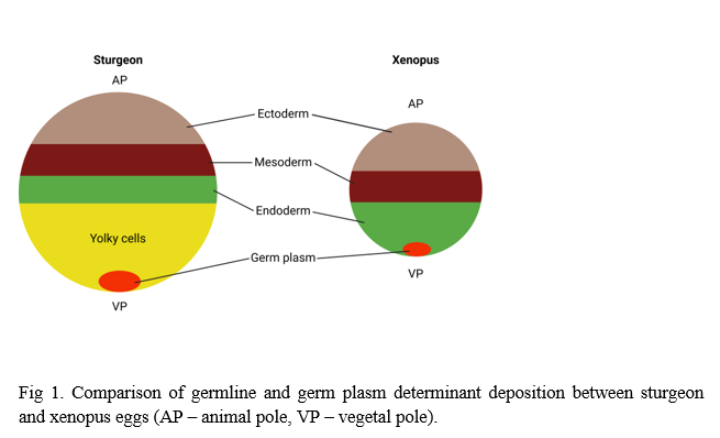
Primordial germ cells (PGCs) in fish are typically specified through maternally inherited germ plasm. In sturgeons (Acipenseridae), this localization is spatially and developmentally distinct, resulting in a unique natural separation of germ cells from somatic lineages during early embryogenesis. Unlike most fish species, sturgeons exhibit holoblastic cleavage and an embryonic development pattern that more closely resembles amphibians rather than typical teleost fishes. This distinctive embryonic architecture establishes sturgeon embryos as a powerful model for studying PGC biology, mitochondrial contributions to germline development, and evolutionary transitions in cleavage patterns.
Methods
We combined PGC visualization using FITC-dextran and GFP-nos3 3’UTR mRNA microinjections, targeted UV irradiation to eliminate vegetal germ plasm, and mitochondrial transplantation experiments in different sturgeon species. Recent findings on the role of vegetal blastomeres and novel techniques for selective blastomere inhibition were integrated. Histological and gene expression analyses (qPCR tomography) were performed to distinguish embryonic and extra-embryonic contributions.
Results
Our data confirmed that sturgeon vegetal blastomeres primarily contribute to PGC formation, while the majority differentiate into transcriptionally inactive yolk cells serving as an extra-embryonic nutritional source. UV irradiation of the vegetal pole effectively ablated PGCs without disrupting somatic development, verifying the independence of early somatic patterning from germ plasm . Mitochondrial transplantation successfully restored PGC formation even when endogenous germ plasm was destroyed, suggesting mitochondria contribute critically to germline specification. Fate-mapping studies indicated that vegetal yolk cells do not integrate into embryonic tissues, supporting a model where sturgeons exhibit an evolutionary transition between holoblastic and meroblastic cleavage.
Conclusion
Sturgeon embryos, with their naturally separated germ plasm and somatic territories, provide an exceptional in vivo system to study fundamental questions of germline development, mitochondrial function, and evolutionary developmental biology in fish. These insights not only advance basic understanding but also have implications for biotechnological applications, including germline conservation strategies for endangered sturgeon species.