A NEW TURBELLARIAN PARASITE INFLICTING SERIOUS MORTALITIES IN RED DRUM AQUACULTURE
Introduction
Turbellarian flatworms are controverted organisms with changing taxonomical adscription. Most of these platyhelminths are terrestrial and aquatic free-living organisms , but also include symbiotic species , and few cases of parasitic ones associated to fish, crustaceans and molluscs . Here, we report an epizootic due to a rhabdocoelan infection in cultured red drum (Sciaenops ocellatum) in a sea- cage farm in the Indian Ocean. We describe the morphological, histological and molecular approaches for its identification.
Methods
Two subsequent outbreak s in 2018-2019 affected red drum in the first year after entering the farm (weight ranging from 12g to 180 g). Water temperature ranged from 23 to 29.5° C and salinity was 36 ‰. Differe nt types of samples of fish presenting acute mortalities and clinical signs were taken. Fresh smears of gills were observed at light microscope on site. Necropsied gills were fixed in 10% neutral buffered formalin (NBF), processed routinely, embedded in Technovit7100- resin, sectioned at 1 µm and stained with Giemsa, PAS, and alcian blue-PAS . Some NBF-fixed specimens were dehydrated, cleared with d imethyl p hthalate and stained with i ron a cetic carmine. A nother sample set was stored in 70% ethanol for molecular identification. The ribosomal genes 18S and 28S were PCR-amplified (Giribet et al., 1996; Jovelin & Justine, 2001) and used concatenated to infer a phylogeny by Bayesian i nference. O ther representatives of Rhabdocoela present in GenBank were included in the analyses . Bacterial samples were taken for basic bacteriological analysis.
Results
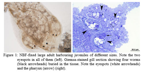
Prevalence of infection was 100 % in some stocks, and mortality ranged from 5% to 60% . Clinical signs included anaemia, weight loss, pale and necrotic gills with mucous masses, desquamation and erosion of the skin , and asphyxiation . Bacteriological results were variable, from negative to opportunistic bacteria or septicaemia, often accompanied by splenomegaly. Microscopical observation of gill scrapings of affected fish revealed ciliated turbellarians with eyespots . When gravid adults were mounted in seawater under a glass coverslip , active swimming eyed juvenile emerged. Worms, with characteristic anterior eyes, were visible under low magnification (Fig. 1). The morphometric study of f ixed worms showed that adults were elongated, piriform with pointed posterior end (854-1403 × 356-589 µm) and a short anterior distal projection . Eyespots were separated. Pharynx was subconical and anterior. Testes and ovary were small and follicular. Some large specimens exhibited numerous completely develop ed juveniles occupying most of the body . Based on the body shape, pharynx arrangement and the presence of juveniles, the worms were tentatively assigned to the family Graffillidae.
H istologically, worms were placed in shallow epithelial tunnels on secondary and primary lamella, and even on the cartilages of gill arches , and some small specimens were found free among gill filaments (Fig.1) . The infection caused destruction of the normal gill architecture with minor histopathological reaction , lacking signs of necrosis or inflammation. Among the epithelial host cells forming the tunnel walls, little or no focal hyperplasia was observed. The primary direct effect of the turbellarian gill infection was the loss of respiratory function by impairment of gas exchange in parasitized lamella. In some sections, long fi lamentous bacteria covered the tissue and worm surface, suggesting secondary bacterial infections involved in the epizootic case. The worms enclosed within epithelial tunnels appeared to be covered by a sheath of cellular and mucous material.
The molecular study placed the parasite within the Order Rhabdocoela, S uborder Dalytyphloplanida and Infraorder Neodalyellida . However, it was highly divergent from all the deposited sequences of the group, indicating that it may belong to a new species not yet described. It did not match with the recent sequence of the old known Pseudografillaria arenicola (Meixner, 1938), and i t could be similar to a turbellarian causing epitozootics (with mortalities > 60%) in the same fish species as well as in other cultured marine fish in China (Wang et al., 2002).
Attempts to treat the infections with formalin baths were unsuccessful.
Conclusions
The histological examination revealed the invasive nature of the worms infecting red drum, and the gill damage could easily explain the anaemia and the asphyxiation of the fish. According to the obtained molecular data, the available orphan sequences and morphological descriptions, t he species could be new to science, but probably present in other far distant locations and hosts . Studies are ongoing for the full description of the species. The current study and the previous reports on turbellarians causing lesions on various marine fish from the Pacific , Caribbean, Chinese and Australian waters, suggest that these parasites may represent and emerging problem in aquaculture, as they are transmitted fish-to-fish, and topic treatments can be ineffective since they live within gill tissues. Future studies are needed to decipher if other reservoir hosts could be involved in its transmission to culture d fish, and which farming conditions favour its blooming.
References
Giribet G., Carranza S. , Baguñà J. , Riutort M., Ribera C. 1996. First molecular evidence for the existence of a Tardigrada + Arthropoda clade. Mol. Biol. Evol., 13: 76-84.
Jovelin R., Justine J.L. 2001 . Phylogenetic relationships within the polyopisthocotylean monogeneans (Platyhelminthes) inferred from partial 28S rDNA sequences. Int . J . Parasitol ., 31: 393-401.
Meixner J. 1938 . Turbellaria (Strudelwürmer ). Die Tierwelt der Nord- und Ostsee . Allgemeiner Teil . In: G. Grimpe , E. Wagler , A Remane (eds). 33(IVb): 1-146, 220 f.
Wang, Y -Y. et al. 2002 . Turbellarian parasitised in Sciaenops ocellatus and its control. J . Fish. China, 26: 379-381.