ONTOGENESIS OF THE RAINBOW TROUT Oncorhynchus mykiss INTESTINE AND ITS STEM CELL NICHE FROM LARVAL TO JUVENILE
Introduction
The transition from endogenous to exogenous feeding characterizes early developmental stages of farmed fish and it is often associated with high mortality rates, resulting in economic loss and reduced animal welfare1,2. The gut and its stem cell niche, as major actors in nutrient absorption and drivers of epithelial development respectively, play a key role in this delicate event3. Therefore, expanding the current understanding of intestinal ontogenesis could improve farming efficiency. In this study, we investigated the intestinal morphological and cellular changes across three early developmental stages of rainbow trout (Oncorhynchus mykiss) - larval, parr, and juvenile – particularly focusing on the cellular populations of the intestinal stem cell niche.
Materials and methods
Whole larvae (20 dph) and i ntestinal samples from parr ( 50 dph ) and juvenile (12 months) rainbow trout were collected and processed for histological purposes . Briefly, we used: (i) h ematoxylin and eosin to assess the general tissue morphology; (ii) Masson’s trichome to highlight the compactum layer of the submucosa; (iii) A lcian blue (pH 2,5) and Periodic Acid Schiff , used in combination, to detect mucins-secreting cells. P roliferating intestinal cells were identified through immunofluorescence of proliferating cell nuclear antigen (PCNA); in situ-hybridization was used to detect the expression of sox9 and pdgfrα as specific markers of intestinal stem cells and telocytes - a stromal cell population regulating stem cell differentiation.
Results
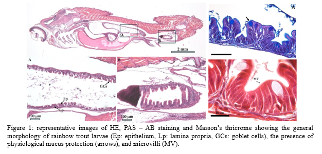
The larval intestine was largely immature: i ts mucosa was organized in rudimental folds whilst the undeveloped submucosa lacked the typical granular cells, later involved in immune defense. However, even at this early developmental stage, the epithelium was characterized by microvilli and mucin-secreting cells, indicating the presence of mucus protection (Fig 1). Epithelial cells proliferation was widespread without a well-defined epithelial stem cell compartment. On the contrary , telocytes already assumed their typical arrangement, creating a complex mesh just below the enterocytes ’ basement membrane. This suggests that telocytes are the first element of the stem cell niche to appear.
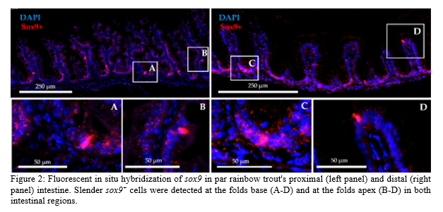
The transition to the parr stage was marked by a more structured morphology with intestinal folds acquiring a finger-like phenotype and an increased complexity of the submucosa . At this stage, a fully developed epithelial stem cells niche restricted to the fold’s base became evident (Fig 2). Moreover, at this stage, enteroendocrine cells appeared scattered along the epithelium.
At the juvenile stage, t he mucosa acquired its definitive configuration with long finger-like folds and a fully developed submucosa consisting of the three distinctive layers: the two granulosum and the thick collagen compactum layer located in between.
Conclusion
The timing of these events suggests that telocytes play a key role in triggering and guiding the formation of a well-defined stem cell niche and that its differentiation is largely completed by the parr stage .
These results expand and integrate the current understanding of the ontogeny of the rainbow trout intestine, providing novel insights into this delicate phase . The definition of the accurate timing of the detailed intestine development may help in optimizing feeding strategies and improving animal welfare, limiting possible economic losses.
Acknowledgements :
This work was supported by NUTRIsim
References
1. Busch, A. Transition from endogenous to exogenous nutrition: Larval size parameters determining the start of external feeding and size of prey ingested by Ruegen spring herring Clupea harengus. Mar. Ecol. Prog. Ser. 130, 39–46 (1996).
2. Wiborg, K. F. Larval mortality in marine fishes and the critical period concept. ICES J. Mar. Sci. 37, 111 (1976).
3. Barker, N. Adult intestinal stem cells: Critical drivers of epithelial homeostasis and regeneration. Nat. Rev. Mol. Cell Biol. 15, 19–33 (2014).
4. Verdile, N., Pasquariello, R., Brevini, T. A. L. & Gandolfi, F. The 3d pattern of the rainbow trout (Oncorhynchus mykiss) enterocytes and intestinal stem cells. Int. J. Mol. Sci. 21, 1–30 (2020).
5. Verdile, N., Pasquariello, R., Cardinaletti, G. & Tibaldi Emilio, Brevini Tiziana A.L., G. F. Telocytes: Active Players in the Rainbow Trout (Oncorhynchus mykiss) Intestinal Stem-Cell Niche. Animals 12(19), 74 (2022).